

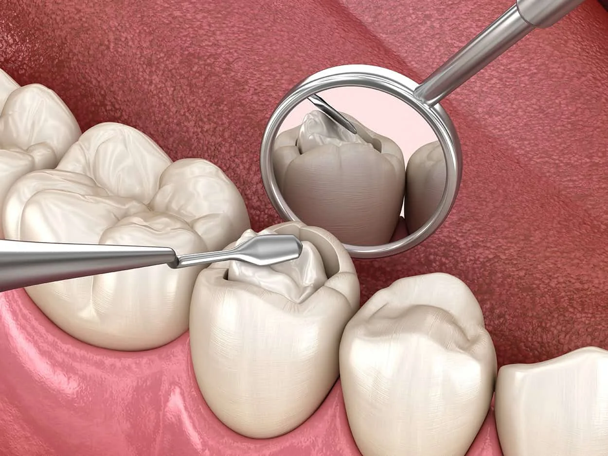
The idea that cavities might be reversible sounds almost too good to be true—but it is not entirely wrong. The accurate version is more nuanced: early decay in a specific stage can be arrested and partially reversed through remineralization. Once a cavity progresses beyond that stage, however, no amount of brushing, fluoride, or dietary change will close it. Understanding the difference is something a local dentist can explain based on what is actually visible on examination and X-rays.
How Decay Actually Develops
The cavity process begins with bacterial acid, not with a hole. Bacteria in the oral biofilm metabolize sugars and carbohydrates and produce acids as a byproduct. Those acids temporarily lower the pH on the tooth surface, dissolving calcium and phosphate ions from the enamel—a process called demineralization. Saliva contains the minerals needed to replenish what was lost and neutralize the acid, so brief demineralization events after eating are a normal part of oral biology that the mouth manages continuously.
Problems arise when the balance tips—when acid attacks are frequent enough, or salivary protection is reduced enough, that demineralization outpaces remineralization over time. The enamel loses net mineral content, becoming increasingly porous and structurally weakened in the affected area. This is the early decay stage. At some point, the enamel surface loses enough integrity that it collapses inward, forming the physical cavity that requires a filling.
The Window When Remineralization Is Actually Possible
Early demineralization that has not yet broken through the enamel surface is the only stage where natural reversal is biologically possible. At this point, the enamel structure is still intact—it is mineral-depleted but not physically breached. Fluoride is the most effective remineralizing agent available because it integrates into the enamel crystal structure during remineralization and produces a harder, more acid-resistant mineral than the original enamel contained.
Clinically, this early decay appears as a white spot on the enamel surface—chalky, opaque, and typically located near the gumline or in between teeth. A local dentist assessing a white spot lesion will evaluate its texture, location, and whether it has progressed to cavitation before recommending a remineralization protocol or a filling. The difference between those two clinical findings is not always visible to the patient, but it is visible to a trained clinician with appropriate lighting and probing.
What Remineralization Actually Requires
Remineralization does not happen passively. Reversing early decay requires reducing the acid challenges that depleted the enamel in the first place while simultaneously providing the mineral supply needed to rebuild it. That means reducing the frequency of sugar and acid exposure in the diet, not just the total amount. Sipping sweetened beverages for hours exposes enamel to continuous acid attack; drinking the same amount in a single sitting gives saliva time to recover between meals.
Fluoride toothpaste used twice daily provides the most accessible source of remineralizing fluoride for most patients. Prescription-strength fluoride, professionally applied fluoride varnish, and remineralizing products containing casein phosphopeptide-amorphous calcium phosphate are used for higher-risk patients or more advanced white spot lesions. The response depends on the severity of the lesion, the patient’s saliva quality and volume, and how consistently the remineralization protocol is followed.
Why Early Decay That Feels Fine Is Still a Problem
Early decay produces no pain. The enamel has no nerve supply of its own, so demineralization in the enamel layer is entirely silent. Many patients assume that because nothing hurts, nothing is happening. In reality, the lesion may be actively progressing through the enamel on its way to the dentin layer, where the presence of nerve-adjacent tubules means pain begins and where decay accelerates significantly.
Dentin is substantially softer than enamel—it decays roughly five times faster. A cavity that has crossed the enamel-dentin junction can progress from a small lesion to a large one in a fraction of the time it took to form in the enamel. This is why early decay that looks minimal on an X-ray today may look dramatically different six months later if nothing is done to address it.
What Happens When Remineralization Is Not Enough
When early decay has progressed past the stage where remineralization can close the defect—when there is a physical cavity in the enamel, decay has entered dentin, or the lesion is in a location that makes home remineralization protocols unreliable—a filling is the appropriate treatment. Fillings do not grow back if left untreated; the bacteria within the cavity continue metabolizing, and the decay front continues advancing toward the pulp.
The size of a filling at the time of placement is directly related to how long treatment was delayed. A small cavity treated promptly requires a small restoration. The same cavity treated six months later may require a larger filling, and a year later may require a crown. The biological process does not pause while the patient waits.
Early Decay Is Manageable—but Only When It Is Found
The truth about early decay is that a narrow window of reversibility exists, and it is identifiable on clinical examination. Outside that window, only a restoration closes the defect. A local dentist who finds a white spot lesion and recommends monitoring with a remineralization protocol is not being cautious—they are responding to a genuinely reversible finding with the least invasive appropriate option.
If you want to learn more about cavity treatment, visit our Local Dentist in Foothill Ranch page or schedule a consultation.